Oral CT Board Review Samples
Below you will find a sampling of oral cardiothoracic surgery board review
questions. Note the variety of formats. Some are short answer with a quick
response, while others require interpretation and synthesis of a variety of
sources of information with a longer answer. Many of the questions include
pearls of wisdom and links to clinical images that you should know for your
exam.
>>
Want More? Sign up today for
a special offer on a board review package. Click Here.
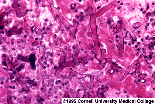
1. Your examiner hands you a color pictograph of the following and asks you for a diagnosis.

What is the diagnosis?
You don't know the answer so he gives you the following clue: it is a PAS stain of a lung wedge biopsy from a diabetic male with pulmonary infiltrates.
It's a PAS stain of an opportunistic lung infection. What is the diagnosis?
-
Who is susceptible to this oportunistic pulmonary infection?
-
What organ systems are affected by this opportunistic pulmonary infection?
-
and What is the treatment of choice for this opportunistic pulmonary infection?
Reference:
Baue AE et al. Glenn's Thoracic and Cardiovascular Surgery, Sixth Edition. Appleton & Lange, Stamford, Conn., 1996 (page 316).
Answer:
Rhizopus, Mucor. These fungi have large, non-septated hyphae with 90 degree angle-branching and non-parallel walls.
Grocott-Gomori methenamine-silver stain is the best stain to use, but hematoxylin and eosin and periodic acid-Schiff (PAS) also may be used. The blood vessels appear thrombosed and hyphae may be seen invading the vessels.
The spores of these fungi are ubiquitous and gain entrance to the body through the mouth and nose. Immunocompetent individuals will phagocytize these spores. Mucormycosis almost exclusively occurs in immunocompromised patients or patients with metabolic abnormalities.
The spores attach to nasal and oral mucosa where massive spore formation occurs. Mucor directly invades blood vessels. Spread occurs when it invades the nasal cavity and maxillary sinuses. Extension to ethmoid sinuses can lead to orbital involvement. Intracranial spread can occur through the ophthalmic artery, superior fissure or cribriform plate. Areas of ischemic infarction and necrosis are seen in the infected tissue. The fungi invade blood vessels and cause thrombosis through inflammatory occlusion.
Fifty to 75% of patients have poorly controlled diabetes mellitus and ketoacidosis. Diabetic patients are predisposed to mucormycosis because of the decreased ability of their neutrophils to phagocytize and adhere to endothelial walls. Furthermore, tissue acidosis and hyperglycemia provide an excellent medium for fungal growth.
Treatment includes therapy with intravenous amphotericin B and aggressive correction of hypoxia, acidosis and hyperglycemia. In addition, aggressive surgical debridement of all necrotic tissue is warranted, sometimes requiring multiple trips to the operating room.
Author's Note: I have known examinees who have been given color pictographs of histology slides to make a diagnosis.
>>
Want More? Sign up today - Click Here.
2.
A. True or False: The attachment of the posterior leaflet of the mitral valve to the annulus has the same length as the free edge of the posterior leaflet.
B. How much (one half? one third? etc.) of the length of the posterior leaflet can be resected in mitral valve repair.
C. How much (one half? one third? etc.) of the length of the FREE EDGE of the anterior leaflet can be resected in mitral valve repair.
Reference:
Antunes MJ. Mitral Valve Repair. Verlag R.S. Schulz, Germany, 1989 (page 84).
Answer:
A. True. Because the attachment of the posterior leaflet of the mitral valve to the annulus has the same length as the free edge of the posterior leaflet, a rectangular (quadrangular) segment of the posterior leaflet can be resected during mitral valve repair.
Links to Further Information & Clinical Images:
Quadrangular Resection of the Posterior Leaflet of the Mitral Valve.
Author Note: A colleague of mine was aksed this particular question on his oral ABTS examination in 2004.
B. Up to one-third of the length of the posterior leaflet can be resected in mitral valve repair. Remember, length of free edge = length of attachment to annulus for the posterior leaflet.
C. Up to one-fifth of the length of the free edge of the anterior leaflet can be resected in mitral valve repair.
>>
Want More? Sign up today - Click Here.
3. A 30 year old male patient with no past medical history undergoes a physical examination for fatigue and is noted to have a continuous murmur on auscultation of the chest.
A. Name two cardiovascular diseases that cause continuous murmurs.
B. Unlike the patent ductus arteriosus, this continuous murmur is heard best during diastole. What is the most likely diagnosis?
C. Are coronary artery fistulae found in patients without congenital heart defects?
D. Coronary artery fistulae are associated with what types of congenital heart diseases?
E. What is the most likely source of a coronary artery fistula?
F. What is the most frequent site of termination of a coronary artery fistula?
G. What is the natural history of coronary artery fistulae?
H. What are indications for closure of coronary artery fistuale?
I. What are the options to close these fistulae?
J. Are patients at risk for developing infective endocarditis?
Reference:
Karamanoukian HL. Cardiac Surgery Board Review. Magalhaes Scientific Press, New York (in press).
Answers:
A. Patent ductus arteriosus and coronary artery fistulae.
B. Coronary artery fistula. The murmur of a coronary artery fistula is typically auscultated lower on the sternal border. Additional features of the coronary artery fistula is diastolic augmentation of the continuous murmur. The ductus arteriosus murmur may have systolic augmentation.
The most likely diagnosis in this case is a coronary artery fistula.
C. Yes. Although they are most often seen in patients with congenital heart defects, they are also found in patients with normal hearts.
D. Right ventricular outflow tract obstruction : pulmonary atresia or stenosis.
E. The right coronary artery is the most likely origin (60%) of a coronary artery fistula.
F. In descending order of frequency, the most likely sources of termination of a coronary artery fistula are : right ventricle, right atrium, coronary sinus, pulmonary artery.
G. Spontaneous closure is rare. Most get bigger over time.
H. Left-to-right shunt (Qp/Qs exceeding 1.5), congestive heart failure and coronary ischemia are indications for closure of coronary artery fistulae. Another important but rare indication is development of infective endocarditis.
I. Options for closure are transcatheter techniques (embolization via coils, etc) or directly using sternotomy and cardiopulmonary bypass. Typically, the the right atrium or pulmonary artery trunk or main branch is opened and the terminal end sutured. If the fistula enters the ventricle or if the feeding vessel is large, the coronary artery is opened, and the origin of the fistula within the coronary artery is closed primarily. This is done to avoid a ventriculotomy.
J. Patients with untreated coronary artery fistulae are at risk for developing infective endocarditis and require prophylaxis with antibiotics for any gastrointestinal, dental or urologic procedures.
>>
Want More? Sign up today - Click Here.
4. Chronic Lung Abscess in a Transplant Patient
A 38 year old kidney transplant patient is admitted for aspiration pneumonia. Although he is treated adequately with intravenous antibiotics for 6 weeks, he is readmitted with fevers, chills and is found to have a thick walled cavity in the chest apex on CT scan with underlying parenchymal lung destruction. There is a crescent shaped radiolucency within this cavity with a central mass that shifts when the patient is placed in the prone position on CT scanning. What is the most likely diagnosis?
Invasive aspergilloma is a life threatening aggressive disease that affects immunocompromised patients. Typical CT abnormalities in pulmonary aspergilloma are a 'halo sign', air crescent lesions, a cavitary lesion or consolidation of the lung associated with pneumothorax.
According to Daly et al., the most common indication for operation was an indeterminate mass, hemoptysis, or severe cough. Lobectomy, wedge excision and pneumonectomy were the most frequent operations. Complications occurred in 78% of patients with complex aspergilloma and in 33% of patients with simple aspergilloma (note: this is consistently tested on ABTS examinations).
They have demonstrated that operative mortality was 5% in patients with simple aspergilloma and 34% in patients with complex aspergilloma (note: this is consistently tested on ABTS examinations).